how do they x ray babies hips
An X-ray of the pelvis focuses specifically on the area between your hips that holds many of your reproductive and digestive organs. In some children with cerebral palsy the femoral head can slowly begin to.

Hip Dysplasia When You Re Too Young For A Hip Replacement Periacetabular Osteotomy Pao Hip Replacement Recovery Hip Replacement Bursitis Hip
The American Academy of Pediatrics does not recommend routine ultrasounds for every infant.

. You will go in the room with him he will need to be stripped from the waist down they will take x-rays of him flat on his back legs dead straight and together you wil be able to hold him in this position then an x-ray of his still on his back with his knees bent facing outwards and the soles of his. Totaleclipse 07092007 1731. Hip ultrasounds take less than 20 minutes and the child will not feel any pain during the examination.
What will happen during the x-ray. This is a basic article for medical students and other non-radiologists. The acetabulum is the socket.
A hip x-ray also known as a hip series or hip radiograph is a pelvis x-ray with an additional lateral view of the specified hip. X-rays can be taken once your baby is 3 months old. Appointments and Referrals.
A hip x-ray also known as a hip series or hip radiograph is a pelvis x-ray with an additional lateral view of the specified hip. No cuts are needed. You may need to leave the room while the pictures are taken.
Its a cast that goes around both hips and down the leg to keep the hips aligned. An X-ray technician will take pictures of the hip. Your baby will be placed on a table and positioned depending on which body area needs an x-ray.
Hip ultrasounds are a safe non-invasive procedure that does not use any radiation. This image shows the soft tissues and the bones of the pelvis and hip joints. A hip X-ray is a safe and painless test that uses a small amount of radiation to make images of the hip joints where the legs attach to the pelvis.
During the examination an X-ray machine sends a beam of radiation through the pelvic bones and hip joints and an image is recorded on a computer or special film. You will go in the room with him he will need to be stripped from the waist down they will take x-rays of him flat on his back legs dead straight and together you wil be able to hold him in this position then an x-ray of his still. Two tests are performed called the Barlow and Ortolani tests to examine the function of the hip joints.
If she does have it they may try to brace it first. If an X-ray of the hip joints is performed according to Launstein Lauenstein then the patients position looks like this. The femoral head should be seated deeply within the acetabulum.
If it persists they may put on a spica cast. Then a surgeon gently pushes the ball of their thighbone joint into the hip socket where it belongs. This is a basic article for medical students and other non-radiologists.
During the examination an X-ray machine sends a beam of radiation through the pelvic bones and hip joints and an image is recorded on a computer or special film. From the front anteroposterior view or AP from the side lateral view also known as the frog leg lateral view Typically X-rays of both hips are taken for comparison even if only one hip is causing symptoms. The first thing to look at in a hip x-ray is the relationship between the femoral head and the acetabulum.
Hip X-rays are done with a child lying on a table. After around 4 to 6 months of age X-rays are the preferred method for evaluating and monitoring hip dysplasia. The rest of your babys body will be covered to protect him or her from the x-ray beam.
The scan can be done on babies up to about 6 months of age. The X-ray image is black and white. The black-and-white images show the internal structures of the hip including the ball-shaped top of the thighbone femoral head and its socket acetabulum in the pelvic bone.
An x-ray can be done on any area of your babys body. An X-ray technician will take pictures of the hip. The hip of the bent limb is pulled aside as far as possible so that the hip joint takes the position of external rotation that is the.
A hip click can be felt by the examiner when the hip joints may not have formed normally. But for babies with an abnormal physical exam or major risk factors for developmental dysplasia of the hip or DDH family history Breech position etc the AAP supports referral for. It is put on by an orthopedic surgeon while using.
It is used for the assessment of unilateral hip pathology most commonly to diagnose a hip fracture or dislocation. Lying on his back one leg in the knee bends at an angle of 30 45 or 90 while its foot rests on the shin of a straightened leg. An ultrasound machine sends sound waves into the hip area and images are recorded on a computer.
Ultrasounds use inaudible sound waves which bounce off of the bones and muscles to create an image for radiologists to interpret. It is used for the assessment of unilateral hip pathology most commonly to diagnose a hip fracture or dislocation. It might help to feed your baby just before the ultrasound to make your little one more relaxed.
Think of the femoral head as the ball of the ball and socket joint.

Degenerative Joint Disease Frog Leg Hip Radiograph Shows Superolateral Joint Space Narrowing Sclerosis Subchondral Cyst A Radiography Osteophyte Radiology

Pin On Radiology Aka X Rays

Pin On X Rays

Anatomy Pathology Medicine Nursing Radiography Radiologictechnologist Radiology Radiologystudent Instagram Medical Anatomy Radiology Student Radiology

Pin On Kallie Mae Pup

Pin On X Ray Imaging

X Ray Image Of Child Swallowed The Coins For A Medical Diagnosis Medicine Pictures Children Images X Ray Images

Pin On Nursing 1st Semester
Radiology Radiologic Imaging Signs List Collection Illustrated Cases Xray X Ray Gi Gu Chest Th Radiology Avascular Necrosis Avascular Necrosis Hip

Froggied Princess Our Life With A Wheaton Pavlik Harness Diapering And The Harness Cloth Hip Babies Diaper Baby Scrapbook

Pin On X Rays

Developmental Dysplasia Of The Hip Ddh Diagnostic Imaging Developmental Dysplasia Of The Hip Diagnostic Imaging Case Study

Anatomy And Physiology Anatomy Sacroiliac

Pin On Pavlik Harness

Pin On Adult Hip Dysplasia Awareness
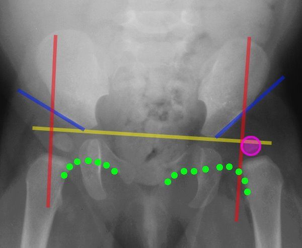
Lines Of The Hip Pediatrics Pediatrics Pediatric Nurse Practitioner Pediatric Radiology

Pin On درمانی

Pin On Hip Dysplasia Pao

Pin By Meg Carter On Ortho Hip Dysplasia X Ray Orthopedics